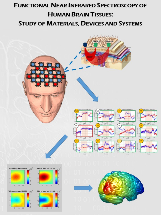
Traditional medical imaging techniques are powerful and have great advantages, but they suffer from some drawbacks limiting their use: ionizing radiations and strong magnetic fields are used as probe but they are risky for health. Moreover the instrumentation forces the patient in a controlled environment or to lay within small space. Near-Infrared spectroscopy (NIRS) and diffuse optical tomography (DOT) are optical techniques that use near-infrared light to probe biological tissues, overcoming the above-mentioned issues. The aim of this study is the deep knowledge of the light behaviour in different inhomogeneous scattering materials, in order to open the way for an extension of fNIRS and DOT techniques from human head or breast to other areas of the human body, such as the chest, characterized by cavities similar to air gaps. In order to increase the performance of these optical techniques, a recently developed category of photodetectors is studied, the Silicon Photomultiplier devices (SiPMs), and a deep characterization of their issues in fNIRS context has been presented in this work. Finally, SiPMs have been used to build a 156-channels fiber-less prototype of an optical system tested on a multilayered optical phantom after its deep characterization, carried out by means of analytical and computational methods (Monte Carlo simulations).

